
Red blood cells are the most common type of blood cell and the vertebrate body's principal means of delivering oxygen to the body tissues via the blood. The cells are filled with hemoglobin, a biomolecule that can bind to oxygen. They take up oxygen in the lungs or gills and release it while squeezing through the body's capillaries. The blood's red color is due to the color of hemoglobin. In humans, red blood cells develop in the bone marrow, take the form of flexible biconcave disks, lack a cell nucleus, subcellular organelles and the ability to synthesize protein, and live for about 120 days.[1]

Red blood cells are also known as RBCs, red blood corpuscles (an archaic term), haematids or erythrocytes (from Greek erythros for "red" and kytos for "hollow", with cyte translated as "cell" in modern usage). The capitalized term Red Blood Cells is the proper name in the US for erythrocytes in storage solution used in transfusion medicine.[2]
Contents[hide]
|
[edit] Vertebrate erythrocytes


Erythrocytes consist mainly of hemoglobin, a complex metalloprotein containing heme groups whose iron atoms temporarily link to oxygen molecules (O2) in the lungs or gills and release them throughout the body. Oxygen can easily diffuse through the red blood cell's cell membrane. Hemoglobin in the erythrocytes also carries some of the waste product carbon dioxide back from the tissues; most of the carbon dioxide is however transported as bicarbonate dissolved in the blood plasma. Myoglobin, a compound related to hemoglobin, acts to store oxygen in muscle cells.[3]
The color of erythrocytes is due to the heme group of hemoglobin. The blood plasma alone is straw-colored, but the red blood cells change color depending on the state of the hemoglobin: when combined with oxygen the resulting oxyhemoglobin is scarlet, and when oxygen has been released the resulting deoxyhemoglobin is darker, appearing bluish through the vessel wall and skin. Pulse oximetry takes advantage of this color change to directly measure the arterial blood oxygen saturation using colorimetric techniques.
The sequestration of oxygen carrying proteins inside specialized cells (rather than having them dissolved in body fluid) was an important step in the evolution of vertebrates; it allows for less viscous blood, higher concentrations of oxygen, and better diffusion of oxygen from the blood to the tissues. The size of erythrocytes varies widely among vertebrate species; erythrocyte width is on average about 25% larger than capillary diameter and it has been hypothesized that this improves the oxygen transfer from erythrocytes to tissues.[4]
The only known vertebrates without erythrocytes are the crocodile icefishes (family Channichthyidae); they live in very oxygen rich cold water and transport oxygen freely dissolved in their blood.[5] While they don't use hemoglobin anymore, remnants of hemoglobin genes can be found in their genome.[6]
[edit] Nucleus
Erythrocytes in mammals are anucleate when mature, meaning that they lack a cell nucleus. In comparison, the erythrocytes of nearly all other vertebrates have nuclei; the only known exception being salamanders of the Batrachoseps genus.[7]
[edit] Functions besides oxygen transport
When erythrocytes undergo shear stress in constricted vessels, they release ATP which causes the vessel walls to relax and dilate.[8]
When their hemoglobin molecules are deoxygenated, erythrocytes release S-nitrosothiols which also acts to dilate vessels,[9] thus directing more blood to areas of the body depleted of oxygen.
Erythrocytes also play a part in the body's immune response: when lysed by pathogens such as bacteria, their hemoglobin releases free radicals that break down the pathogen's cell wall and membrane, killing it.[10][11]
[edit] Mammalian erythrocytes
Mammalian erythrocytes have nuclei during early phases of development, but extrude them as they mature in order to provide more space for hemoglobin. Mammalian erythrocytes also lose their other organelles such as their mitochondria. As a result, the cells use none of the oxygen they transport; instead they produce the energy carrier ATP by fermentation, via glycolysis of glucose followed by lactic acid production. Furthermore, red cells do not have an insulin receptor and thus their glucose uptake is not regulated by insulin. Because of the lack of nuclei and organelles, mature red blood cells do not contain DNA and cannot synthesize any RNA, and consequently they cannot divide or repair themselves.
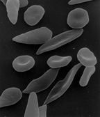
Mammalian erythrocytes are biconcave disks: flattened and depressed in the center, with a dumbbell-shaped cross section. This shape (as well as the loss of organelles and nucleus) optimizes the cell for the exchange of oxygen with its surroundings. The cells are flexible so as to fit through tiny capillaries, where they release their oxygen load. Erythrocytes are circular, except in the camel family Camelidae, where they are oval.
In large blood vessels, red blood cells sometimes occur as a stack, flat side next to flat side. This is known as rouleaux formation, and it occurs more often if the levels of certain serum proteins are elevated, as for instance during inflammation.
The spleen acts as a reservoir of red blood cells, but this effect is somewhat limited in humans. In some other mammals such as dogs and horses, the spleen sequesters large numbers of red blood cells which are dumped into the blood during times of exertion stress, yielding a higher oxygen transport capacity.

[edit] Human erythrocytes
A typical human erythrocyte disk has a diameter of 6–8 µm and a thickness of 2 µm, much smaller than most other human cells.[12] A normal erythrocyte has a volume of about 90 fL. About a third of the volume of Red Blood Cells is made up of hemoglobin. A total of 270 million hemoglobin molecules, with each carrying four heme groups is contained in each Red Blood Cell.
Adult humans have roughly 2–3 × 10¹³ red blood cells at any given time (women have about 4 to 5 million erythrocytes per microliter (cubic millimeter) of blood and men about 5 to 6 million; people living at high altitudes with low oxygen tension will have more). Red blood cells are thus much more common than the other blood particles: there are about 4,000–11,000 white blood cells and about 150,000–400,000 platelets in each microliter of human blood.
In humans, hemoglobin in the red blood cells is responsible for the transport of more than 98% of the oxygen; the remaining oxygen is carried dissolved in the blood plasma.
The red blood cells of an average adult human male store collectively about 2.5 grams of iron, representing about 65% of the total iron contained in the body.[13][14] (See Human iron metabolism.)
[edit] Life cycle
The process by which red blood cells are produced is called erythropoiesis. Erythrocytes are continuously produced in the red bone marrow of large bones, at a rate of about 2 million per second. (In the embryo, the liver is the main site of red blood cell production.) The production can be stimulated by the hormone erythropoietin (EPO), synthesised by the kidney; this is used for doping in sports. Just before and after leaving the bone marrow, the developing cells are known as reticulocytes; these comprise about 1% of circulating red blood cells.
Erythrocytes develop from committed stem cells through reticulocytes to mature erythrocytes in about 7 days and live a total of about 100-120 days.
The aging erythrocyte undergoes changes in its plasma membrane, making it susceptible to recognition by phagocytes and subsequent phagocytosis in the spleen, liver and bone marrow. Much of the important breakdown products are recirculated in the body. The heme constituent of hemoglobin are broken down into Fe3+ and biliverdin. The biliverdin is reduced to bilirubin, which is released into the plasma and recirculated to the liver bound to albumin. The iron is released into the plasma to be recirculated by a carrier protein called transferrin. Almost all erythrocytes are removed in this manner from the circulation before they are old enough to hemolyze. Hemolyzed hemoglobin is bound to a protein in plasma called haptoglobin which is not excreted by the kidney.
[edit] Membranes and surface proteins
The membranes of red blood cells play many roles that aid in regulating immune recognition and deformability.
There are two main types of proteins on the surface:
- Band 3
- Glycophorins such as glycophorin C
The blood types of humans are due to variations in surface glycoproteins of erythrocytes.
Disorders of the proteins in these membranes are associated with many disorders, such as hereditary spherocytosis, hereditary elliptocytosis, hereditary stomatocytosis, and paroxysmal nocturnal hemoglobinuria.
[edit] Separation and blood doping
Red blood cells can be obtained from whole blood by centrifugation, which separates the cells from the blood plasma. During plasma donation, the red blood cells are pumped back into the body right away and the plasma is collected. Some athletes have tried to improve their performance by blood doping: first about 1 litre of their blood is extracted, then the red blood cells are isolated, frozen and stored, to be reinjected shortly before the competition. (Red blood cells can be conserved for 5 weeks at −79 °C.) This practice is hard to detect but may endanger the human cardiovascular system which is not equipped to deal with blood of the resulting higher viscosity.
[edit] Artificially grown red blood cells
In 2008 it was reported that human embryonic stem cells had been successfully coaxed into becoming erythrocytes in the lab. The difficult step was to induce the cells to eject their nucleus; this was achieved by growing the cells on stromal cells from the bone marrow. It is hoped that these artificial erythrocytes can eventually be used for blood transfusions.[15]
[edit] Diseases and diagnostic tools
Blood diseases involving the red blood cells include:
- Anemias (or anaemias) are diseases characterized by low oxygen transport capacity of the blood, because of low red cell count or some abnormality of the red blood cells or the hemoglobin.
- Iron deficiency anemia is the most common anemia; it occurs when the dietary intake or absorption of iron is insufficient, and hemoglobin, which contains iron, cannot be formed
- Sickle-cell disease is a genetic disease that results in abnormal hemoglobin molecules. When these release their oxygen load in the tissues, they become insoluble, leading to mis-shaped red blood cells. These sickle shaped red cells are rigid and cause blood vessel blockage, pain, strokes, and other tissue damage.
- Thalassemia is a genetic disease that results in the production of an abnormal ratio of hemoglobin subunits.
- Spherocytosis is a genetic disease that causes a defect in the red blood cell's cytoskeleton, causing the red blood cells to be small, sphere-shaped, and fragile instead of donut-shaped and flexible.
- Pernicious anemia is an autoimmune disease wherein the body lacks intrinsic factor, required to absorb vitamin B12 from food. Vitamin B12 is needed for the production of hemoglobin.
- Aplastic anemia is caused by the inability of the bone marrow to produce blood cells.
- Pure red cell aplasia is caused by the inability of the bone marrow to produce only red blood cells.
- Hemolysis is the general term for excessive breakdown of red blood cells. It can have several causes.

- The malaria parasite spends part of its life-cycle in red blood cells, feeds on their hemoglobin and then breaks them apart, causing fever. Both sickle-cell disease and thalassemia are more common in malaria areas, because these mutations convey some protection against the parasite.
- Polycythemias (or erythrocytoses) are diseases characterized by a surplus of red blood cells. The increased viscosity of the blood can cause a number of symptoms.
- In polycythemia vera the increased number of red blood cells results from an abnormality in the bone marrow.
- Several microangiopathic diseases, including disseminated intravascular coagulation and thrombotic microangiopathies, present with pathognomonic (diagnostic) RBC fragments called schistocytes. These pathologies generate fibrin strands that sever RBCs as they try to move past a thrombus.
- Hemolytic transfusion reaction is the destruction of donated red blood cells after a transfusion, mediated by host antibodies, often as a result of a blood type mismatch.
Several blood tests involve red blood cells, including the RBC count (the number of red blood cells per volume of blood) and the hematocrit (percentage of blood volume occupied by red blood cells). The blood type needs to be determined to prepare for a blood transfusion or an organ transplantation.
[edit] History
The first person to describe red blood cells was probably the young Dutch biologist Jan Swammerdam, who had used an early microscope in 1658 to study the blood of a frog.[16] Unaware of this work, Anton van Leeuwenhoek provided another microscopic description in 1674.[17]
No comments:
Post a Comment